New review paper: exploring how CNS phagocytes orchestrate neuroinflammation

We are excited to share that our new review paper is featured on the cover of the March issue of Trends in Neurosciences!

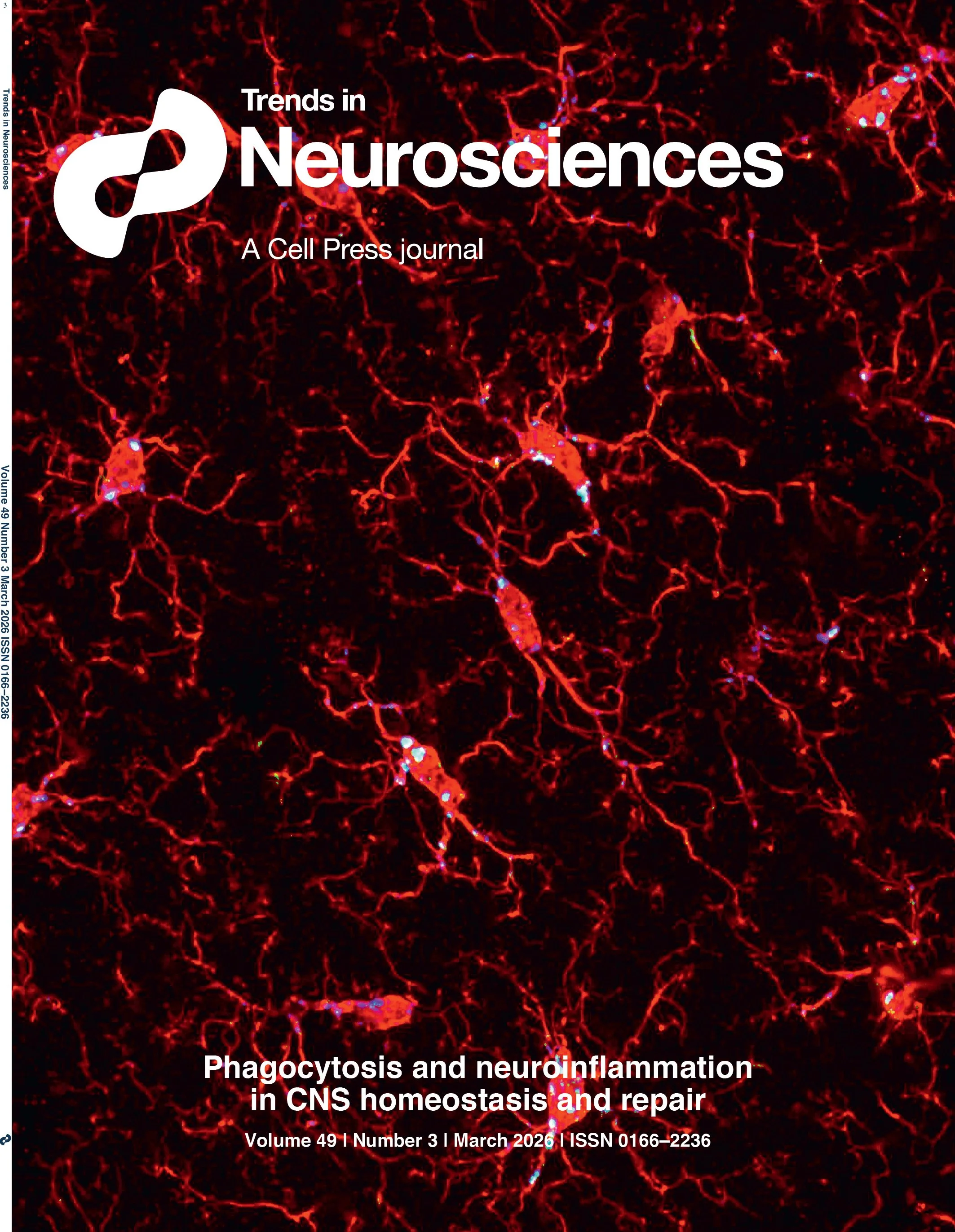
Trends in Neurosciences cover, March 2026 - Gaudet and Fonken. The cover shows hippocampal microglia (red, Iba1) with engulfed postsynaptic material (green, PSD95) in phagolysosomes (blue, CD68) during development. Image credit: Brandy Routh, Fonken lab.

The paper, “Phagocytosis and neuroinflammation: orchestrating CNS homeostasis, repair, and the resolution of inflammation,” was a collaboration with Laura Fonken (UT Austin College of Pharmacy). We developed a framework to describe how phagocytosis - the cellular ingestion of debris - crucially regulates the inflammatory response in the brain and spinal cord.

Beyond Cleanup: Phagocytosis as an Inflammatory Conductor

Phagocytosis is often oversimplified as a passive cleanup process. However, the molecules that drive debris clearance also dictate the inflammatory tone of the CNS. In our review, we explore how this double-edged sword impacts various pathologies:

  • Homeostasis: Microglia and astrocytes manage synapse pruning and apoptotic cell removal to maintain health.

  • Disease and injury: The role of receptors like TREM2 and TAM receptors in clearing myelin debris in MS, amyloid-β in Alzheimer’s, and necrotic tissue after CNS injury and stroke.

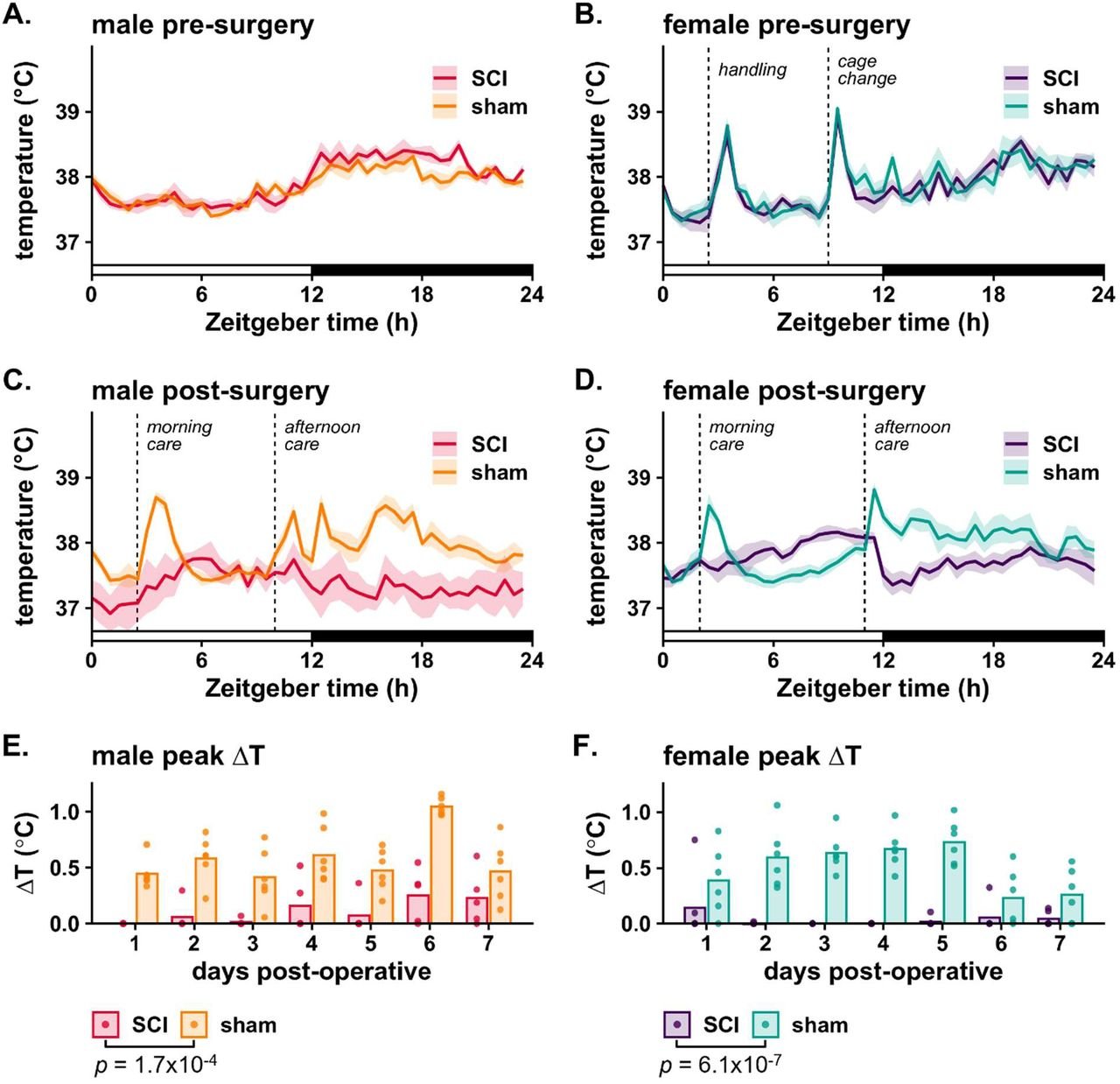
  • Regulating debris removal and inflammatory state: We discuss how activating phagocytic systems can drive removal of damaging debris, while also shaping pro- versus anti-inflammatory cell state. These cell states have implications for disease and repair trajectories.

  • Complexities with "eat-me" signals: We highlight the delicate balance in the peri-lesion environment, where phagocytes must distinguish between toxic debris and viable, but stressed, neurons.

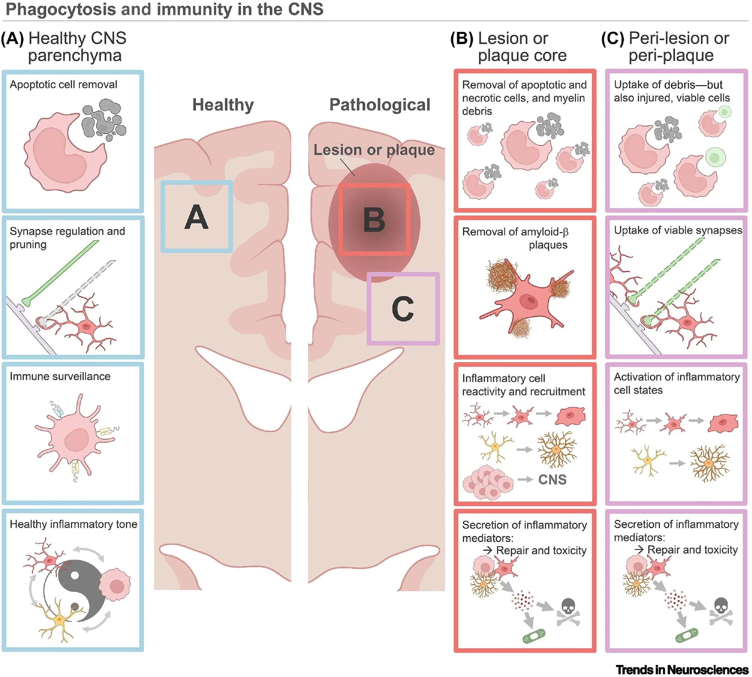
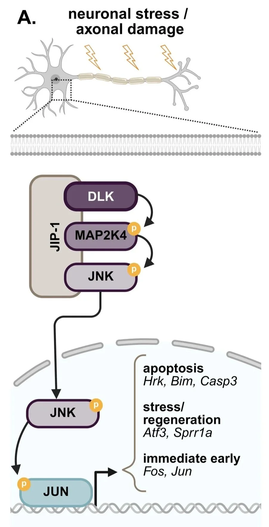
Figure 1: Phagocytosis and immunity in the central nervous system.

Future Therapeutic Directions

A major focus of the review is the transition from understanding mechanisms to fine-tuning these pathways to enhance recovery. We discuss emerging strategies - including TREM2 agonists, bispecific antibodies, and focused ultrasound - designed to optimize debris clearance while protecting healthy tissue.

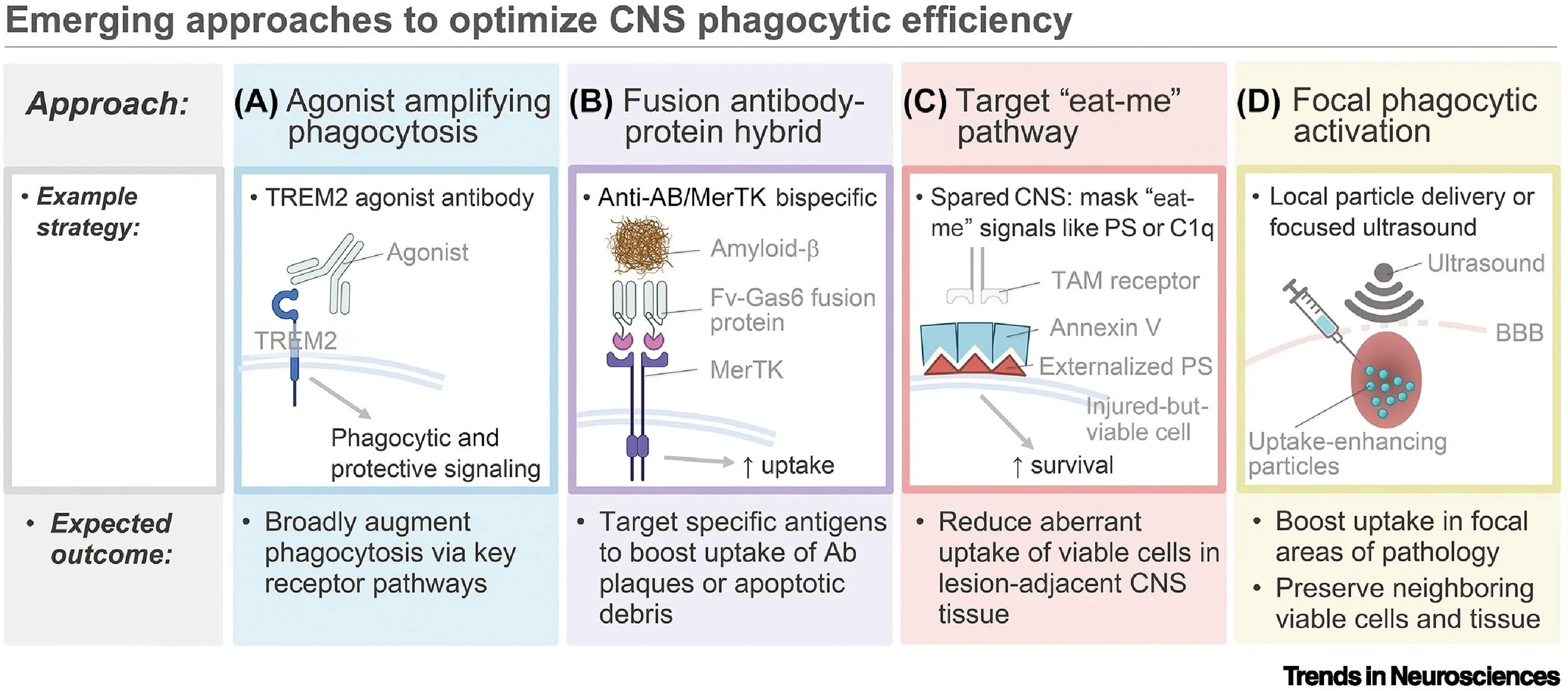
Figure 3 from the paper: Emerging approaches to optimize CNS phagocytic efficiency.

We are grateful to the editors at Trends in Neurosciences for the cover feature and the opportunity to synthesize these evolving concepts. We hope this framework helps guide future research into these phagocytic regulators that resolve neuroinflammation and promote repair.

Check out the full study here: https://authors.elsevier.com/a/1mi1vbotqBbQu (temporary free link)
DOI: https://doi.org/10.1016/j.tins.2026.01.002

Authors: Andrew D. Gaudet and Laura K. Fonken

The DLK dilemma: Why a "death signal" might be necessary after spinal cord injury

Spinal cord injury (SCI) involves primary trauma, followed by a wave of “secondary damage” - an acute process involving radiating inflammation and cellular stress, killing neurons that survived initial impact. At the Gaudet Lab, we are searching for molecular modulators that control this process, hoping to shift inflammation to benefit tissue survival and functional recovery.

Figure 1A from the paper: Schematic of the DLK signaling cascade. Extracellular stress activates DLK, which phosphorylates MAP2K4, leading to phosphorylation of JNK. JNK translocates to the nucleus and activates the transcription factor JUN.

Our latest research, “DLK inhibition has sex-specific effects on neuroprotection and locomotor recovery after spinal cord injury” (pdf here), focused on a protein called Dual Leucine Zipper Kinase (DLK). In other models of neurodegeneration, like glaucoma or Alzheimer’s, DLK acts as a primary stress sensor. When a neuron is damaged, DLK sends a "death signal" through the JNK pathway (a molecular messenger system), telling the cell to shut down (Fig. 1 from the paper). We hypothesized that if we could block DLK and this death signal immediately after a spinal cord injury, we could shield these vulnerable neurons and help mice regain more movement.

To test this, we used a selective DLK inhibitor (IACS-52825) in a mouse model of spinal cord contusion. This work was led by Dr. John Aldrich (a Research Scientist in the lab), in collaboration with Dr. W. Jim Ray and other experts at the Belfer Neurodegeneration Consortium at the University of Texas MD Anderson Cancer Center. Using single-nucleus RNA sequencing, we confirmed that the DLK pathway is activated in neurons for at least a week following injury. In addition, the drug effectively entered the spinal cord and successfully suppressed the stress-activated signals.

However, the functional results took us by surprise.

A Surprising Sex Difference

Interestingly, our results contradicted our prediction. Instead of improving recovery, inhibiting DLK made outcomes worse after SCI - but only in males.

Figure 6 from the paper: DLK inhibitor increased lesion size in males, but not females after SCI. (A) In males (left), treatment with the DLK inhibitor led to significantly larger lesion areas (the central gap) compared to controls. (B,C) Quantification of lesion area and volume in males and females.

As shown in Figure 6, male mice treated with the inhibitor had 71% larger SCI lesions. This worsened damage was associated with a delay in their ability to regain coordinated walking. The female mice told a completely different story; the drug had no significant effect on their lesion size or their locomotor recovery after SCI (as seen in Figure 5).

Figure 5C and E from the paper: DLK inhibitor worsened open field locomotor recovery in males, but not females after SCI.

IMPLICATIONS

These results underscore why we must study both sexes in medical research. In the male spinal cord, the DLK "stress signal" might be beneficial in the early days post-injury - perhaps helping to coordinate the initial immune response or priming neurons for repair. By blocking DLK, we may have inadvertently disrupted a necessary part of the body's natural repair process. Further, broad inhibition of DLK likely impacts various cell types, including both neurons and oligodendrocytes. DLK also likely performs different functions in different parts of the cell. Optimizing how we target DLK - considering specific cell types, locations, and timing - could still lead to effective therapies for SCI and other neurologic conditions.

Check out the full study here:DLK inhibition has sex-specific effects on neuroprotection and locomotor recovery after spinal cord injury” (pdf)

Authors (Gaudet lab members bolded):
John C. Aldrich, Samantha M. Alman, Sydney E. Lee, Ashley R. Scheinfeld, Chelsea C. Zhang, Averi L. Pike, Fiona C. Bremner, Olivia Calderon, Sunil Goodwani, William J. Ray, Andrew D. Gaudet

Gaudet visits the University of Maryland STAR Center

On December 8th, Dr. Gaudet visited the Center for Shock, Trauma and Anesthesiology Research (STAR) at the University of Maryland School of Medicine. It was a privilege to present our lab's recent work on neuroinflammation and recovery to such a dedicated group of trauma researchers. A huge thank you to Dr. Junfang Wu for being a wonderful host and for the insightful discussions on new approaches for studying neuroinflammation and supporting trainees.

Mission Connect Symposium: connecting for a cure in Houston

On November 21st, the Gaudet Lab headed to Houston for the annual Mission Connect Symposium. It was a fantastic day of networking and sharing ideas with the Texas neurotrauma community. A highlight of the event was the keynote by Dr. Jae K. Lee (University of Miami), whose work on the molecular mechanisms of spinal cord injury repair continues to inspire our own research. We’re grateful to Mission Connect and the TIRR Foundation for organizing such a vibrant event to push SCI research forward.

G-lab attends the Texas Society for Circadian Biology and Medicine in Dallas

The Gaudet Lab traveled to Dallas on November 8th for the 2025 Texas Society for Circadian Biology and Medicine (TSCBM) meeting. We have a long-standing interest in how biological rhythms impact neuroinflammation and neurobiology, so it was an honor to be surrounded by the giants of the field. Our team experienced remarkable comments with Nobel Laureate Dr. Michael Rosbash and Dr. Joseph Takahashi, whose pioneering work discovered the Clock gene. It was an energizing day of chronobiology and new ideas!

Discussing research at the University of Mississippi Medical Center

Our fall seminar tour kicked off on September 23rd at the University of Mississippi Medical Center (UMMC). Dr. Gaudet had the pleasure of presenting our latest findings on sex differences and SCI recovery to the faculty and students. Special thanks to Dr. Dobrivoje (Boba) Stokic and Dr. Matthias Krenn for the invitation and the warm hospitality. Jackson had incredible historic neighborhoods with superb dining and a beautiful capitol building. UMMC is a remarkable center with excellent clinical and training environments! The visit was a great opportunity to discuss the intersection of basic science and clinical rehabilitation.

New research support: targeting chronic pain with funding from TRC4

We are excited to announce that the Gaudet Lab has received a new research grant from the Trauma Research and Combat Casualty Care Collaborative (TRC4). This funding will support our work on a major hurdle in recovery: chronic neuropathic pain following peripheral nerve injury.

While current pain medications often have detrimental side effects like sedation or the risk of addiction, our team is investigating a more precise molecular target: PKCε (protein kinase C epsilon). PKCε is a key "switch" in peripheral sensory neurons that drives the transition from acute to chronic pain.

In collaboration with Dr. Robert Messing and Dr. Stanton McHardy, we are testing a new small molecule (CP612) designed to inhibit PKCε. Our goal is to determine whether blocking this pathway can dampen persistent pain and improve functional recovery after peripheral nerve injury.

By focusing on the molecular drivers of pain, we hope to develop better therapeutic options for both veterans and civilians recovering from trauma. We are appreciative to receive support from TRC4 and look forward to sharing our progress!

Ashley and Andrew presented new science at Neurotrauma 2025 in Philadelphia!

The Neurotrauma 2025 Symposium was a blast! It was great to connect with colleagues over science and culinary adventures in Philadelphia.

Ashley Scheinfeld - a graduate student in the lab - presented her latest compelling findings regarding the role of phagocytic TAM receptors in controlling neuroinflammation, recovery, and pain after SCI. Great work, Ashley! We’re excited to see where your research leads.

Andrew led an insightful plenary session on neuroinflammation after traumatic brain injury and spinal cord injury with the brilliant Drs. Ona Bloom, Ruchira Jha, and David Loane. The talks thoughtfully linked preclinical and clinical ideas and elicited creative discussions. The audience size and engagement was impressive - particularly because the session was bright and early after a fun karaoke night!

Also, it was great to experience Philadelphia for the first time. Reading Market and downtown had innumerable delicious food options - and we were a short run away from the Art Museum and the Rocky statue.

Thanks to all the speakers, organizers, and attendees for making NNS2025 such a memorable and thought-provoking event. We’re looking forward to next year's conference in Milwaukee!

Celebrating progress with pizza!

Today was a great day in the lab! We gathered for a delicious pizza lunch to celebrate recent successes and enjoy some time socializing together.

It was also a wonderful opportunity to recognize Chelsea Zhang and Kalina Dusenbery as they prepare for their next big steps. Chelsea will soon be heading off to Jefferson University to start medical school, and Kalina will be starting graduate studies here at UT-Austin!

We're incredibly proud of both of them. Congratulations, Chelsea and Kalina - we wish you all the best on your exciting journeys ahead!

Postbac student Kalina Dusenbery won a prestigious Harrington Fellowship to attend grad school at UT-Austin!

Congratulations to Kalina Dusenbery, our incredible postbac student, who has been awarded the highly competitive Harrington Fellowship to pursue her graduate studies here at UT-Austin!

Kalina is passionate about the circadian system and its complex connections to the CNS and immunity. We're thrilled that she will continue exploring these fascinating links, joining the Fonken lab here at UT. Her research will help illuminate how our internal clocks affect CNS and immune function.

Congrats, Kalina!

Uapii Kandjoze joins the Gaudet lab as a new graduate student!

We're thrilled to announce that Uapii Kandjoze has officially joined our lab as a graduate student!

Uapii is a student in the highly regarded Interdisciplinary Neuroscience Program here at UT-Austin. She earned her Bachelor's degree in Neuroscience from Earlham College in Indiana. Uapii has already excelled in previous research positions at Case Western Reserve University and Drexel, where she explored important neuroscience topics including Alzheimer's disease models, blood-brain barrier integrity, and epilepsy.

In the Gaudet lab, Uapii will bring her expertise to our work on spinal cord injury, focusing on novel approaches to relieve pain and improve motor function.

Welcome, Uapii!

Spinal cord injury's hidden impact: disrupting your body's stress thermostat

Imagine facing a stressful situation – maybe a thunderstorm quickly approaching or a surprise encounter on a hiking trail. Your body gears up, heart racing, senses heightened. Part of that healthy stress response often includes a subtle rise in core body temperature, preparing you for action.

For individuals with spinal cord injury (SCI), this natural response can be dramatically altered. SCI severely impacts the autonomic nervous system, which controls involuntary body functions like heart rate, blood pressure, and digestion. This disruption can lead to serious conditions like autonomic dysreflexia, where the body overreacts dangerously to stimuli.

Despite these widespread autonomic issues, how SCI affects the body's ability to control temperature, especially during stress, has been largely overlooked. Our latest preprint addresses this gap, revealing in rats how SCI affects crucial temperature-related stress responses.

Female and male rats were implanted with a small transmitter that measured activity and core temperature. Two weeks later, rats received T8 contusion SCI or sham laminectomy surgery.

Next, we assessed body temperature across the day in rats prior to and after SCI (or sham) surgery (Fig. 1). Prior to surgery, both male and female rats exhibit expected daily rhythms in core temperature, with higher temperatures during the active (dark) phase. We noted that handling elicited a stress response, including increased activity and body temperature. In sham-surgery rats, this stress-induced hyperthermia is maintained immediately after surgery. In contrast, SCI rats lack stress-elicited hyperthermia in the acute phase after SCI. This SCI-driven loss of stress-induced hyperthermia occurs in both female and male rats.

Figure 1. The stress-induced hyperthermic response is abolished acutely after SCI.

(A,B) Average core temperature (± SEM) for male (A) and female (B) rats is shown at 1-2 weeks pre-surgery. The x-axis represents Zeitgeber Time (ZT), with ZT0 denoting lights on and ZT12 lights off. Handling/care events are annotated with vertical dashed lines. (C,D) Sham and SCI temperatures at 2 dpo – whereas sham rats show typical stress-induced hyperthermia, SCI rats lose this hyperthermic response. (E,F) Temperature peaks above background (ΔT) for sham and SCI rats for 7 dpo, with bars representing group means and points marking individual values. SCI disrupts stress-induced hyperthermia across the acute post-injury period. P-values, indicating significant main effects of surgery, were determined via mixed-effects model.

Remarkably, this temperature control deficit was not permanent (Fig. 2). Over several weeks post-injury, both male and female rats with SCI gradually regained their ability to mount a stress-induced hyperthermia response. Interestingly, females with SCI recovered their stress responses slightly faster than males.

Figure 2. Stress-induced hyperthermia is abolished over acute-to-subacute times after incomplete SCI, and is regained by chronic post-SCI times.

(A,B) Heatmaps display ΔT (temperature peaks above background) for individual male (A) and female (B) rats across 7 weeks post-surgery. The first care or interaction event of the day is shown. Cage changes are marked with “v”. Solid lines indicate twice-daily care (1–17 dpo for males and 1–14 for females), while dotted lines mark once-daily care (27 dpo for males and 28 dpo for females). Days with no care events or missing data are shaded in grey. (C,D) Modeled recovery curves for the stress-induced hyperthermic response in male and female animals are based on daily estimated marginal means derived from a mixed-effects model. Lines represent the modeled estimates ± standard error. Vertical lines indicate the estimated stress-hyperthermic recovery dates, defined as the first day when SCI stress-induced hyperthermia responses were statistically indistinguishable from sham responses (male SCI vs. male sham: 27 dpo; female SCI vs. female sham: 23 dpo) (p > 0.05). (E,F) Average core temperatures (± SEM) are shown for sham/SCI males and females on the estimated stress-induced hyperthermia recovery date (left panel, E and F) and during the final cage change at 7 weeks post-surgery (right panel). Room entries for cage changes are annotated with vertical dashed lines.

Our findings reveal a significant, and transient, disruption of temperature regulation and stress-induced hyperthermia following T8 SCI. This research opens new opportunities for understanding the broader impact of SCI on the autonomic nervous system. Ultimately, by uncovering and addressing these critical physiological deficits, we aim to help develop interventions that could improve the quality of life for individuals living with SCI.

This exciting work includes contributions by John Aldrich, Kalina Dusenbery, Linda Watkins, and Andrew Gaudet. We appreciate support provided by University of Texas at Austin start-up funds, the Wings for Life Foundation (Watkins/Gaudet), and NIH NINDS R01NS131806 (Gaudet).

Andrew presents new research at the Winter Brain Conference

Andrew Gaudet recently shared exciting new data from the lab at the thought-provoking Winter Brain Conference in beautiful Lake Tahoe, CA.

Andrew presented during a fascinating session titled, "Pain and spinal cord injury: Underlying mechanisms and impact on recovery," led by Kip Kramer. The session also featured compelling talks by Michelle Hook and Paulina Scheuren. Andrew highlighted some of our latest findings in his presentation, "Sex differences in pain and anxiety after spinal cord injury.”

Beyond science, it was fantastic to connect – and even ski with – friends and colleagues surrounded by such spectacular scenery.

We're already looking forward to seeing more familiar faces at the Neurotrauma Symposium in June!

Welcome, med student intern Chelsea!

We’re excited to welcome Chelsea Zhang to the lab for the next five months!

Chelsea recently completed her Bachelor’s degree at Penn State University, and will be entering medical school in the Fall at Jefferson University. Chelsea will gain valuable research experience in this internship as she prepares for med school.

We’re delighted to have her join us and look forward to seeing what she can achieve in her time here at UT-Austin!

New G-lab paper: Revealing sex differences in anxiety in mice using a novel light-heat conflict test (TIDAL)

We published a paper, titled “Anxiety-like behaviors in mice unmasked: revealing sex differences in anxiety using a novel light-heat conflict test,” in Journal of Neuroscience Research! (pdf here) The study was led by a former graduate student from the lab, Dr. Sydney Lee.

Anxiety is twice as prevalent in women, yet sex differences for anxiety-like behaviors are not detected in rodents using commonly used tests.

In this paper, we develop a new test with two chambers that places an anxiety-inducing stimulus—light—in conflict with heat. The dark chamber floor heats to uncomfortable temperatures, whereas the illuminated chamber temperature remains comfortable. The test is called the Thermal Increments Dark-Light (TIDAL) conflict test.

Our data reveal that females remain on the dark-heating plate for longer than males, suggesting that females display increased anxiety-like behavior.

These anxiety-like behaviors in females were reduced using an anxiety-relieving drug, paroxetine. Since paroxetine reduced mouse preference for the dark-heated plate, this supports the premise that the TIDAL conflict test is a valid test for anxiety-like behavior.

Study summary: Tests for mouse anxiety-related behavior do not reliably recapitulate sex differences in human anxiety. The TIDAL conflict test comprises one dark plate that heats to uncomfortable temperatures, and one illuminated-isothermic plate. Female mice persist on the dark-heated plate longer than males; thus, TIDAL reveals clinically-relevant sex differences in anxiety-related behavior.

Therefore, the new TIDAL conflict test unmasks sex differences in mouse anxiety-like behavior - female mice displayed higher anxiety-like behavior than males, which recapitulates sex differences in anxiety observed in humans. Thus, the TIDAL conflict test could help identify new sex-specific mechanisms and treatments for anxiety.

Our prior work used the TIDAL conflict test to reveal that spinal cord injury in female and male mice induces robust anxiety-like behavior (pdf here). Thus, in addition to unveiling sex-specific mechanisms of anxiety, the TIDAL conflict test will be useful for exploring the relative salience of anxiety vs. heat sensitivity under pathological conditions - e.g., sickness, aging, or neurologic disorders.

Authors on the paper include Sydney Lee, Sung-Hoon Park, John Aldrich, Laura Fonken, and Andrew Gaudet. We appreciate support provided by University of Texas at Austin start-up funds, the Wings for Life Foundation, and Mission Connect, a program of the TIRR Foundation. Research was also supported by NIH NINDS R01NS131806 (Gaudet), and by NIH R01AG062716 (Fonken) and R01AG078758 (Fonken).

Gaudet lab presents at the Annual Mission Connect Symposium in Houston

The Gaudet lab took a road trip to Houston to attend the Mission Connect Annual Symposium. Mission Connect, a program of the TIRR Foundation, is an incredible resource for spinal cord injury researchers in Texas - they hold grant competitions, monthly meetings, and an annual symposium. This year, we were excited to hear from the plenary speaker, Dr. Jeffrey Twiss. We stayed well caffeinated, and trainees received constructive feedback on their posters. We appreciate this excellent meeting, and look forward to seeing everyone again in Houston in 2025!

Gaudet and Fonken lab fun at the Annual Meeting of the Society for Behavioral Neuroendocrinology

The G-lab was represented well in Columbus, OH at the 28th Annual Meeting of the SBN! Kalina Dusenbery presented new data revealing the benefits of a REV-ERB agonist on neuroinflammation after spinal cord injury. The Gaudet and Fonken labs had fun mingling with other behavioral neuroscientists - including at Ohio Stadium! We’re excited to explore this project further, and hope to see SBN colleagues next year in Vancouver, BC.

Gaudet Lab attends Neurotrauma 2024 in San Francisco!

Ashley and Andrew enjoying the poster session.

Ashley and Andrew attended Neurotrauma 2024! Ashley presented her poster on the role of phagocytic receptors in controlling repair after CNS injury. It was a thought-provoking conference with fantastic friends and colleagues. We shared discussion, lunches, and happy hour around the Hilton Union Square in San Francisco.

Lovely afternoon for a run to Golden Gate Beach!

Streetcar at Union Square.

Fonken-Gaudet labs attend the Texas Society for Circadian Biology and Medicine

The Fonken-Gaudet labs took a Saturday trip to Texas A&M and College Station to experience a riveting series of sessions exploring circadian biology. The annual Texas Society for Circadian Biology and Medicine provides a rich forum for learning about chronobiology and meeting other circadian researchers. Texas is so large and has so many research institutes that each of these “small” meetings has expertise that meets or exceeds that found in most countries! We had fun learning about diverse topics and biological systems linked by a shared passion for biological clocks. See you all next year!